Call: 08071931179
 Send Inquiry
 Send Inquiry
Grad Dental OPG And Dental X-Ray Machine
MOQ : 1 Unit
Grad Dental OPG And Dental X-Ray Machine Specification
- Material
- Metal
- Condition
- New
- Technology
- Digital OPG and Cephalometric X-ray
- Portable
- No
- Light Source
- High-frequency X-Ray Tube
- Pressure Range
- Atmospheric
- Operating Temperature
- +10C to +40C
- Humidity %
- 80% RH
- Sensors Specification
- Compatible with digital and analog sensors
- Indicator Specification
- LCD Digital Display Panel
- Darkness Range
- Adjustable Exposure Parameters
- Ray Frequency Range
- 50/60 Hz
- Exposure Time Range (in Sec)
- 0.4 - 3.2 sec
- Focus
- 0.5 mm focal spot
- Image System
- Digital / Film-based option
- Usage
- Hospital
- Power Source
- Electric
- Power Consumption
- 1.5 kW
- Working Voltage
- 220-240V AC, 50/60Hz
- Dimension (L*W*H)
- 1920 mm x 1230 mm x 970 mm
- Weight
- Approx. 160 kg
- Rotation Angle
- 270 panoramic arm rotation
- Tube Current
- 4-10 mA (adjustable)
- Tube Voltage
- 60-90 kV (adjustable)
- Safety Features
- Automatic exposure control, fail-safe mechanism
- Patient Positioning
- Chin rest, bite block and head support
- Beam Type
- Collimated X-ray beam
- Exposure Modes
- Child, Adult, TMJ, Sinus, Bitewing
- Cephalometric Arm
- Optional attachment available
- Color
- White & Blue standard finish
- Mounting
- Floor-mounted, free-standing unit
Grad Dental OPG And Dental X-Ray Machine Trade Information
- Minimum Order Quantity
- 1 Unit
- Payment Terms
- Cash Advance (CA)
- Supply Ability
- 10 Units Per Month
- Delivery Time
- 20 Days
- Main Domestic Market
- All India
About Grad Dental OPG And Dental X-Ray Machine
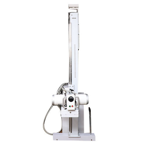
Being a well-distinguished firm in the market, our company is engaged in manufacturing, exporting and supplying the Grad Dental OPG And Dental X-Ray Machine. It is a type of diagnostic imaging equipment used to capture images of the jaws, teeth (basically of the entire mouth). This helps to diagnose the impacted teeth, gum disease, cavities and other dental abnormalities. The offered machines are tested on several parameters to ensure the functioning, durability and performance. Furthermore, the range of the Grad Dental OPG And Dental X-Ray Machine can be availed in different specifications and at nominal prices.
Advanced Imaging and Versatile Functions
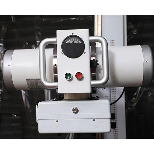
This dental X-ray unit delivers high-quality panoramic and cephalometric images with adjustable exposure parameters for child, adult, TMJ, sinus, and bitewing modes. A 0.5 mm focal spot ensures sharp results, while the system accommodates both digital and film-based workflows. Its robust metal build, digital display panel, and high-frequency X-ray tube make it reliable for busy hospital settings.
Precision Positioning and Patient Comfort
Grad Dental OPG provides ergonomic features such as a chin rest, bite block, and head support to guarantee optimal patient positioning and minimize movement. These positioning aids, combined with a 270 arm rotation, allow for thorough imaging of all dental regions, ensuring both patient comfort and superior diagnostic accuracy.
Safety and Ease of Operation
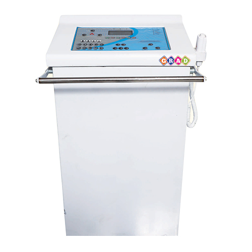
Equipped with automatic exposure control, collimated X-ray beam, and a fail-safe mechanism, this device prioritizes patient and operator safety. The LCD digital display panel simplifies operation and status monitoring. Its simple electric power source and adjustable parameters provide flexibility and quick adaptation for various clinical requirements.
FAQ's of Grad Dental OPG And Dental X-Ray Machine:
Q: How do I operate the Grad Dental OPG and Dental X-Ray Machine for different patient types?
A: The machine offers dedicated exposure modes for child, adult, TMJ, sinus, and bitewing imaging. Select the appropriate mode and adjust tube voltage (60-90 kV) and tube current (4-10 mA) using the LCD digital display. Position the patient securely using the chin rest, bite block, and head support, then proceed with the desired imaging workflow.Q: What benefits does the optional cephalometric arm attachment offer?
A: The optional cephalometric arm provides the capability to capture lateral and posteroanterior skull images, which are essential for orthodontic assessments, treatment planning, and craniofacial analyses. This feature adds versatility, making the system suitable for a broader range of diagnostic applications.Q: When should the adjustable exposure parameters be changed?
A: Exposure parameters, such as tube voltage, current, and exposure time (0.4-3.2 seconds), should be adjusted based on patient age, size, the area being examined, and diagnostic requirements. Higher exposure might be necessary for adults or dense anatomical areas, while pediatric patients require lower settings to ensure safety.Q: Where can this X-ray unit be installed, and what are its mounting requirements?
A: This floor-mounted, free-standing unit is designed for installation in hospital dental imaging suites or dedicated X-ray rooms. It requires a flat, stable surface and a standard power supply of 220-240V AC. Adequate room dimensions should accommodate the machine (1920 mm x 1230 mm x 970 mm) and provide space for panoramic arm rotation.Q: What is the process for switching between digital and film-based imaging?
A: The system is compatible with both digital and analog sensors. To switch, select the appropriate sensor, and connect it to the machine as specified in the manual. Digital images are processed immediately, while film-based imaging requires standard post-exposure development procedures.Q: How does the machine enhance safety during operation?
A: It incorporates automatic exposure control to prevent excessive radiation, a fail-safe mechanism for error protection, and a collimated X-ray beam to restrict exposure only to the imaging area. These features minimize risks for both patients and operators.Q: What maintenance is required to ensure optimal performance?
A: Routine maintenance includes regular calibration, checking safety systems, cleaning the exterior surfaces, and inspecting connections. Follow the manufacturer's guidelines for specific timelines and service procedures to ensure ongoing reliability and diagnostic precision.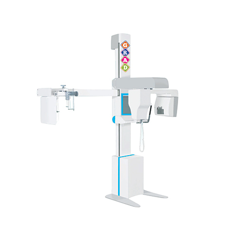
Tell us about your requirement

Price:
Quantity
Select Unit
- 50
- 100
- 200
- 250
- 500
- 1000+
Additional detail
Mobile number
Email

More Products in X-Ray Machine Category
X Ray Tube Head
Minimum Order Quantity : 1 Unit
Condition : New
Portable : Other, No (wall/ceiling mounted or fixed installation)
Usage : Medical diagnostic imaging, dental Xray applications
Material : Aluminum alloy housing with lead shielding
500Ma Static X-Ray Machine
Price 200000 INR / Unit
Minimum Order Quantity : 1 Unit
Condition : New
Portable : No
Usage : Hospital
Material : Metal
Digital Display Mobile X-Ray Unit
Price 120000 INR / Unit
Minimum Order Quantity : 1 Unit
Condition : New
Portable : Yes
Usage : Hospital
Material : Metal
Grad Fix Digital X-Ray Machine
Price 200000 INR / Unit
Minimum Order Quantity : 1 Unit
Condition : New
Portable : Yes
Usage : Hospital
Material : Metal
Contact Details
GRAD MEDICAL EQUIPMENTS PRIVATE LIMITED
GST : 09AAJCG5553E1ZH
- E-93, Sector -7,Noida - 201301, Uttar Pradesh, India
- Phone :08071931179
- Direct Number: 919910735480
Developed and Managed by Infocom Network Private Limited.
